What is a CT?
Computed Tomography (CT) imaging, also referred to as a computed axial tomography (CAT) scan, involves the use of rotating X-ray equipment, combined with a digital computer, to obtain images of the body. Using CT imaging, cross sectional images of body organs and tissues can be produced. CT imaging can provide views of soft tissue, bone, muscle and blood vessels, without sacrificing clarity.
What is CBCT?
Cone Beam Computed Tomography (CBCT) is a compact, faster and safer version of the regular CT. Through the use of a cone shaped X-Ray beam, the size of the scanner, radiation dosage and time needed for scanning are all dramatically reduced.
A typical CBCT scanner can fit easily into any dental ( or otherwise ) practice and is easily accessible by patients. The time needed for a full scan is typically under one minute and the radiation dosage is upto a hundred times less than that of a regular CT scanner.
Uses of CBCT
- To Detect Cancers CT imaging is commonly used for diagnostic purposes. In fact, it is a chief imaging method used in diagnosing a variety of cancers, including those affecting the lungs, pancreas and liver. Using CT imaging, not only can physicians confirm that tumors exist, but they can also pinpoint their locations, accurately measure the size of tumors and determine whether or not they've spread to neighbouring tissues. In addition to the diagnosis of certain cancers, CT imaging is used for planning and administering radiation cancer treatments, as well as for planning certain types of surgeries. It is useful for guiding biopsies and a range of other procedures categorized as minimally invasive.
- CT imaging is a valuable tool for the diagnosis and treatment of musculoskeletal disorders and injuries. It is often used to measure bone mineral density and to detect injuries to internal organs.
- CT imaging is even used for the diagnosis and treatment of certain vascular diseases that, undetected and untreated, have the potential to cause renal failure, stroke, or death.
- A very important tool for planning Implant placements as a CBCT gives accurate analysis of quality and quantity of bone, proximity to vital organs like nerves, sinuses, etc.
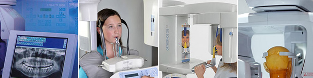
OPG (Orthopantomogram)
What is an orthopantomogram?An orthopantomogram is an panoramic X-ray image of your whole mouth, including your upper and lower jaw and teeth. The X-ray machine moves around your head while taking the image. This provides a complete ear to ear image of your mouth and teeth.
It is useful in following areas:
- Impacted wisdom teeth diagnosis and treatment planning - the most common use is to determine the status of wisdom teeth, their position, their proximity to nerves and trauma to the jaws.
- Periodontal bone loss and periapical involvement.
- Finding the source of dental pain.
- Assessment for the placement of dental implants.
- Orthodontic assessment pre and post operative.
- Caries detection especially in the inter-dental region.
- Diagnosis of developmental anomalies such as cherubism, cleido cranial dysplasia.
- Carcinoma (cancer) in relation to the jaws.
- Temporomandibular joint dysfunctions and ankylosis.
- Diagnosis of osteosarcoma, ameloblastoma, renal osteodystrophy affecting jaws and hypophosphatemia.
- Diagnosis and pre- and post-surgical assessment of oral and maxillofacial trauma, e.g. dentoalveolar fractures and mandibular fractures.
- Salivary stones.
- Any abnormal growths, etc.
Preparing for your orthopantomogram
Before the procedure can take place, you will need to remove glasses, dentures and any jewellery from your head and neck (such as earrings and necklace) as well as any hairclips.
Advantage of panoramic images:
- Broad coverage of facial bones and teeth.
- Low patient radiation dose.
- Convenience of examination for the patient (films need not be placed inside the mouth).
- Particularly invaluable for patients with gagging (vomiting) reflex.
- Ability to be used in patients who cannot open the mouth or when the opening is restricted e.g.: due to trismus.
- Short time required for making the image.
- Patient's ready understandability of panaromic films, making them a useful visual aid in patient education and case presentation.
- Easy to store compared to the large set of intra oral x-rays which are typically used.
What are the risks?
Orthopantomograms are commonly performed and generally safe. However, in order to make an informed decision and give your consent, you need to be aware of the possible risks. You will be exposed to some X-ray radiation but the amount you receive isn't considered to be harmful. The level of exposure will depend on the procedure. Talk to your doctor or radiologist for more information. Pregnant women are advised not to have X-rays as there's a risk the radiation may harm their unborn baby. If you are or could be, pregnant then please tell your doctor or radiographer.